
HEALTH
The Importance Of Nuclear Imaging In The Medical Sector
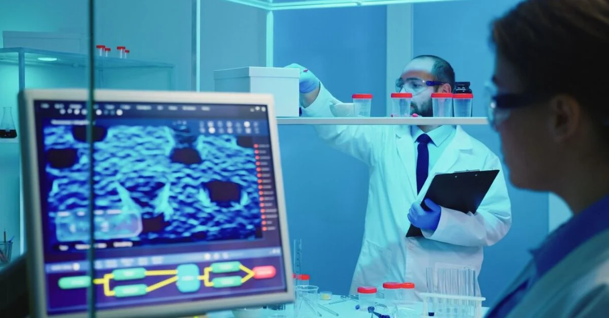
Nuclear imaging is a key diagnostic tool used in healthcare. This advanced technology allows doctors to see inside the body in unique ways. As per experts like PRP Imaging, it helps detect and evaluate many medical conditions. Keep reading to learn more about how nuclear imaging works and why it’s so valuable for patients.
What Is Nuclear Imaging?
Nuclear imaging uses radioactive materials called radiotracers. These tracers release energy. Special cameras detect this energy and create pictures of the body’s interior. This lets doctors view organs and tissues. It shows problems without needing surgery. Two common types of nuclear imaging are PET scans and SPECT scans.
How a PET Scan Works
PET stands for positron emission tomography. The patient gets an injection of a radiotracer. Areas where the tracer collects light up when scanned. This creates 3D images. PET scans are useful for seeing metabolic activity. This means how the body’s cells are functioning. They’re often used to detect cancers.
How a SPECT Scan Works
SPECT is single-photon emission computerized tomography. It’s similar to PET scans. But only one gamma ray photon is emitted from the radiotracer. The 3D images show how blood flows to tissues and organs. SPECT helps evaluate heart and brain disorders. It also assists in cancer detection.
Key Benefits for Patients
Here are some top ways nuclear imaging improves medical care:
Finds disease in earliest stages when treatment works best
- Detects whether cancers have spread to other sites
- Guides biopsy by pinpointing suspicious spots
- Assesses tumors without removing them
- Maps organ function throughout the body
- Checks effectiveness of treatments over time
- Avoids exploratory surgery in many cases
- Provides information unavailable through other tests
- Quick and painless for patients versus more invasive tests
Analyzing Cancer
One major use of nuclear imaging is detecting and analyzing cancers. PET and SPECT provide unique information compared to CT and MRI scans. They show cellular function. This reveals how aggressive tumors are. It spots recurrences and spread earlier than other modalities. Radiotracers identify optimal biopsy locations. Scans indicate how well treatment is progressing.
Evaluating Heart Conditions
Nuclear imaging also excels at assessing heart health issues. SPECT scanning creates detailed 3D maps of heart function and blood flow. This helps diagnose coronary artery disease. It also evaluates damage after a heart attack. PET scans measure blood flow and oxygen levels in the cardiac muscles. This information is vital when planning treatments.
Examining Neurologic Function
In the brain, PET and SPECT track circulation and activity patterns. This is useful after strokes, seizures, concussions and more. Scanning helps diagnose memory disorders like Alzheimer’s. It also assists with epilepsy surgical planning and therapy selection. Dopamine imaging gauges Parkinson’s disease progression.
The Future of Nuclear Imaging
Researchers are developing new radiotracers for expanded applications. Hybrid scanners combining PET and MRI offer enhanced images. As technology improves, nuclear imaging becomes increasingly valuable. It provides doctors invaluable views inside the body. This leads to earlier disease detection plus better treatment planning and follow up.

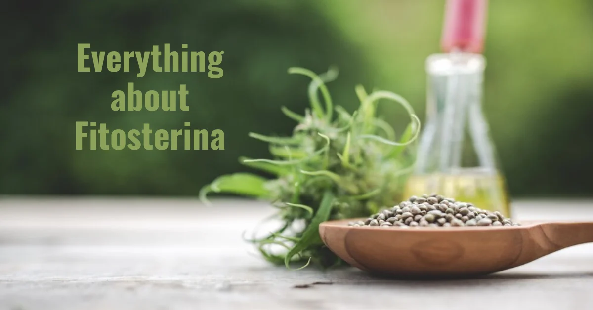
Introduction to Fitosterina
In recent years, there has been growing interest in natural compounds and their potential health benefits. One such compound that has gained attention is Fitosterina. This article aims to explore the various health benefits associated with Fitosterina, its sources, and how it can be incorporated into a healthy lifestyle.
What is Fitosterina?
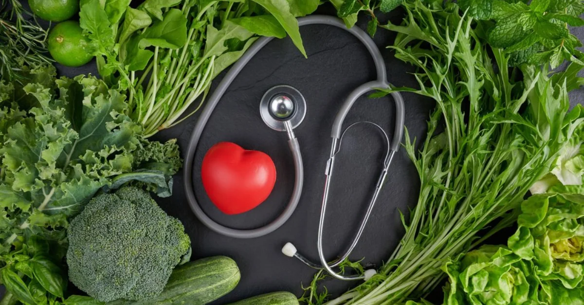
Fitosterina, also known as plant sterols, refers to a group of naturally occurring compounds found in plants. These compounds are structurally similar to cholesterol and are known to have various health benefits, particularly in managing cholesterol levels and promoting heart health.
Sources of Fitosterina
Fitosterina can be found in various plant-based foods such as fruits, vegetables, nuts, and seeds. Some of the richest sources include avocados, almonds, sesame seeds, and olive oil. Consuming a diet rich in these foods can help increase Fitost,erina intake.
Health Benefits of Fitosterina
Lowering Cholesterol Levels:
Fitosterina has been extensively studied for its ability to lower LDL cholesterol levels, also known as “bad” cholesterol. By blocking the absorption of cholesterol in the intestines, Fitost,erina helps reduce overall cholesterol levels in the body, thus lowering the risk of heart disease.
Anti-Inflammatory Properties:
Studies have shown that Fitosterina possesses anti-inflammatory properties, which can help reduce inflammation in the body. Chronic inflammation is linked to various health conditions such as heart disease, diabetes, and arthritis, making Fitost,erina a valuable compound in promoting overall health.
Potential in Cancer Prevention:
Some research suggests that Fitost,erina may have potential in preventing certain types of cancer, including breast and prostate cancer. Although more studies are needed to confirm these findings, preliminary research is promising.
Improving Heart Health:
In addition to lowering cholesterol levels, Fitosterina has been shown to improve overall heart health by reducing the risk of atherosclerosis and coronary artery disease. Including Fitosterina-rich foods in your diet can contribute to better heart health and reduced risk of cardiovascular events.
Managing Diabetes:
Fitosterina may also play a role in managing diabetes by improving insulin sensitivity and reducing blood sugar levels. Including Fitost,erina-rich foods as part of a balanced diet can help individuals with diabetes better manage their condition.
ALSO READ: EVERYTHING ABOUT GLÚTEM
Research Studies on Fitosterina
Numerous research studies have been conducted to investigate the health benefits of Fitosterina. These studies have consistently shown positive results, particularly in relation to cholesterol management, heart health, and inflammation reduction. However, more research is needed to fully understand the mechanisms of action and potential long-term effects of Fitost,erina.
How to Incorporate Fitosterina into Your Diet
Incorporating Fitosterina into your diet is simple and can be done by including a variety of plant-based foods in your meals. Some easy ways to increase Fitost,erina intake include:
- Adding nuts and seeds to salads, yogurt, or smoothies.
- Cooking with olive oil or avocado oil instead of butter or margarine.
- Snacking on avocado toast or guacamole.
- Incorporating whole grains such as oats, barley, and quinoa into your meals.
By making these small dietary changes, you can significantly increase your Fitost,erina intake and reap its health benefits.
Potential Side Effects and Risks
While Fitosterina is generally considered safe for most people when consumed in normal dietary amounts, excessive intake may lead to potential side effects such as digestive issues or interference with the absorption of fat-soluble vitamins. It is essential to consume Fitost,erina as part of a balanced diet and consult with a healthcare professional if you have any concerns or pre-existing health conditions.
ALSO READ: PAINSLTUBE: REDEFINING PAIN RELIEF
Consulting a Healthcare Professional
Before making any significant changes to your diet or lifestyle, it is always advisable to consult with a healthcare professional, especially if you have any underlying health conditions or are taking medications. A healthcare provider can provide personalized recommendations based on your individual health needs and help you incorporate Fitost,erina into your overall wellness plan safely.
Conclusion
In conclusion, Fitosterina is a natural compound found in plant-based foods that offers a wide range of health benefits, including lowering cholesterol levels, reducing inflammation, and improving heart health. By incorporating Fitost,erina-rich foods into your diet and consulting with a healthcare professional, you can optimize your health and reduce the risk of various chronic diseases.
ALSO READ: ALEVEMENTE: WELLNESS WOVEN INTO MIND, BODY, AND SOUL
FAQs
Is Fitosterina safe for everyone to consume?
While Fitost,erina is generally safe when consumed in dietary amounts, individuals with certain health conditions or who are taking specific medications should consult with a healthcare professional before increasing their intake.
Can Fitosterina supplements be beneficial?
Fitost,erina supplements are available, but it’s essential to use them cautiously and under the guidance of a healthcare professional. Whole foods sources of Fitost,erina are preferred, as they come with additional nutrients and are less likely to cause side effects.
How much Fitosterina should I consume daily for optimal health benefits?
There is no specific recommended daily intake for Fitos,terina, but including a variety of Fitosterina-rich foods in your diet regularly can contribute to overall health and well-being.
Are there any interactions between Fitosterina and medications?
Fitost,erina supplements may interact with certain medications, including cholesterol-lowering drugs and blood thinners. It’s crucial to discuss any potential interactions with your healthcare provider before starting Fitos,terina supplementation.
Can Fitosterina help with weight loss?
While Fitos,terina may indirectly support weight loss by promoting heart health and reducing inflammation, it is not a direct weight loss aid. A balanced diet and regular exercise remain the cornerstones of weight management.
Fitosterina, also known as phytosterols, are naturally occurring compounds found in plants. They are structurally similar to cholesterol and play vital roles in plant cell membranes. In recent years, fitosterina has garnered attention for its potential health benefits, particularly in managing cholesterol levels and promoting overall well-being.
What is Fitosterina?
Fitosterina, derived from the Greek word “phyto,” meaning plant, and “sterol,” refers to a group of plant sterols and stanols. These compounds are found in various plant-based foods, such as fruits, vegetables, nuts, seeds, and grains. Fitosterina molecules resemble cholesterol in their structure but differ in their absorption and metabolism within the body.
Sources of Fitosterina
Common food sources rich in fitosterina include:
- Nuts and seeds (e.g., almonds, sunflower seeds)
- Vegetable oils (e.g., olive oil, sesame oil)
- Whole grains (e.g., oats, barley)
- Legumes (e.g., beans, lentils)
- Fruits and vegetables (e.g., avocados, spinach)
Health Benefits of Fitosterina
Lowering Cholesterol
One of the most well-known benefits of fitosterina is its ability to lower LDL (bad) cholesterol levels. Fitosterina competes with cholesterol for absorption in the intestines, leading to reduced cholesterol absorption and lower blood cholesterol levels. Regular consumption of fitosterina-rich foods or supplements can help manage cholesterol levels and reduce the risk of heart disease.
Anti-inflammatory Properties
Fitosterina also exhibits anti-inflammatory properties, which can help alleviate inflammation in the body. Chronic inflammation is linked to various health conditions, including heart disease, diabetes, and certain cancers. By reducing inflammation, fitosterina’s may contribute to overall health and well-being.
Potential Cancer-Fighting Effects
Some research suggests that fitosterina’s may have potential cancer-fighting effects. Studies have shown that fitost,erina may inhibit the growth of cancer cells and induce apoptosis (cell death) in certain types of cancer. However, more research is needed to fully understand the mechanisms underlying fitosterina’s anticancer properties.
ALSO READ: DISCOVERING QUETAQUENOSOL: A JOURNEY TO VITALITY
Fitosterina in Nutrition
Incorporating fitost,erina into the diet is relatively easy, as many plant-based foods naturally contain these compounds. Including a variety of fitost,erina-rich foods in your daily meals can help reap the health benefits associated with these plant sterols.
Recommended Daily Intake
The recommended daily intake of fitost,erina varies, but consuming around 2 grams per day has been suggested to help lower cholesterol levels effectively. However, it’s essential to consult with a healthcare professional before starting any new dietary supplements, especially if you have existing health conditions or are taking medications.
Fitosterina Supplements
For those unable to obtain sufficient fitost,erina from dietary sources alone, supplements are available. Fitosterina supplements typically come in the form of capsules or soft gels and are often derived from plant sources such as soybeans or pine trees.
Dosage and Safety Considerations
When taking fitosterina supplements, it’s essential to follow the recommended dosage instructions provided by the manufacturer. Excessive intake of fitost,erina supplements may lead to adverse effects such as digestive issues or interference with the absorption of fat-soluble vitamins. Pregnant or breastfeeding women should consult with their healthcare provider before taking fitost,erina supplements.
ALSO READ: DEȚ DECODED: A JOURNEY TO MIND-BODY HARMONY
Fitosterina and Heart Health
Impact on Cardiovascular Health
Several studies have demonstrated the beneficial effects of fitosterina on cardiovascular health. By lowering LDL cholesterol levels, fitost,erina helps reduce the risk of atherosclerosis (hardening of the arteries) and coronary artery disease. Including fitosterina-rich foods as part of a heart-healthy diet can contribute to overall cardiovascular wellness.
Fitosterina in Skincare
Benefits for Skin Health
In addition to its internal health benefits, fitosterina also offers advantages for skin health when applied topically. Fitost,erina has moisturizing and anti-inflammatory properties, making it beneficial for dry or irritated skin. Skincare products containing fitost,erina can help nourish the skin and maintain its natural barrier function.
Beauty Products Containing Fitosterina
Fitosterina is a common ingredient in various beauty and skincare products, including creams, lotions, and serums. These products aim to hydrate the skin, reduce inflammation, and improve overall skin texture and appearance. Incorporating fitost,erina-based skincare products into your daily routine can help promote healthy, radiant skin.
ALSO READ: UNDERSTANDING IMMATURE FRUITS AND WHITE SKIN
Risks and Side Effects
While fitosterina is generally considered safe for most people, there are some risks and potential side effects to be aware of.
Possible Adverse Effects
- Digestive issues: Some individuals may experience digestive discomfort, such as bloating or diarrhea, when consuming large amounts of fitosterina-rich foods or supplements.
- Interference with vitamin absorption: Fitost,erina may interfere with the absorption of fat-soluble vitamins (A, D, E, K) when consumed in high doses, leading to nutrient deficiencies over time.
- Allergic reactions: In rare cases, allergic reactions to fitosterina supplements may occur, leading to symptoms such as itching, swelling, or difficulty breathing.
Precautions to Take
- Consult with a healthcare professional before starting fitost,erina supplements, especially if you have underlying health conditions or are taking medications.
- Stick to recommended dosage guidelines to avoid potential side effects.
- Monitor your intake of fitost,erina-rich foods and supplements, especially if you have a history of digestive issues or allergies.
Conclusion
In conclusion, fitosterina, or phytosterols, are plant-based compounds with numerous health benefits. From lowering cholesterol levels to reducing inflammation and potentially fighting cancer, fitost,erina plays a crucial role in promoting overall health and well-being. By incorporating fitost,erina-rich foods into your diet and considering supplementation when necessary, you can harness the power of these plant sterols to support your health goals.
ALSO READ: TRANSFORM YOUR LIFE WITH WELLHEALTH AYURVEDIC HEALTH TIPS
FAQs
Is fitosterina safe for everyone to consume?
While fitost,erina is generally safe for most people, individuals with certain health conditions or allergies should consult with a healthcare professional before incorporating fitost,erina supplements into their routine.
Can fitosterina supplements help lower cholesterol levels?
Yes, fitost,erina supplements have been shown to effectively lower LDL (bad) cholesterol levels when taken as directed. However, it’s essential to follow recommended dosage guidelines and consult with a healthcare provider if you have existing health conditions or are taking medications.
Are there any natural sources of fitost,erina?
Yes, fitost,erina is naturally found in various plant-based foods, including nuts, seeds, vegetable oils, whole grains, fruits, and vegetables. Including these foods in your diet can help increase your fitost,erina intake.
What are the potential side effects of fitosterina supplementation?
Some potential side effects of fitost,erina supplementation include digestive issues, interference with vitamin absorption, and allergic reactions. It’s essential to follow recommended dosage guidelines and monitor for any adverse reactions.
Can fitost,erina benefit skin health?
Yes, fitost,erina offers several benefits for skin health, including moisturizing properties and anti-inflammatory effects. Skincare products containing fitost,erina can help hydrate the skin and improve overall skin texture and appearance.
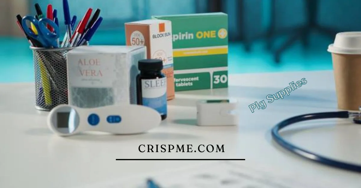
Introduction to PLG Supplies
In a world where innovation and efficiency reign supreme, the concept of PLG supplies has emerged at the forefront of industries ranging from agriculture to healthcare. But what exactly does “PLG” stand for? Product-Led Growth is not just a buzzword; it’s a transformative approach reshaping how businesses operate and engage with their customers. With an emphasis on creating exceptional products that drive user satisfaction, this model impacts everything from farm tools to medical essentials.
As we dive deeper into the realm of PLG supplies, you’ll discover how these components can elevate your business strategy and enhance customer experiences. Whether you’re farming in rural landscapes or navigating the complexities of modern medicine, understanding PLG supplies could be your key to unlocking unprecedented growth opportunities. Join us as we explore this dynamic landscape!
Understanding Product-Led Growth and its Impact on the Business World
Product-Led Growth (PLG) is a strategy that prioritizes the product itself as the main driver of customer acquisition, retention, and expansion. Companies adopting PLG focus on delivering exceptional user experiences directly through their offerings.
This approach shifts the traditional sales funnel. Instead of relying heavily on marketing or sales teams to push products, businesses allow users to discover value independently. By leveraging free trials or freemium models, customers can personally engage with the product before committing financially.
The impact on the business world has been significant. Companies embracing PLG often see quicker scaling and reduced customer acquisition costs. They cultivate stronger relationships with clients since users are more invested in a product they have explored firsthand.
As these companies grow, they learn from user interactions too. This feedback loop helps refine features and enhance overall satisfaction, fostering loyalty in an increasingly competitive landscape.
The Role of Farm Tools in PLG Supplies
Farm tools are essential components of PLG supplies. They empower agricultural businesses to enhance productivity while focusing on customer needs.
Product-led growth thrives in environments where user-friendly innovations shine. Modern farm tools, such as smart irrigation systems and precision seeders, exemplify this trend. These tools simplify operations, making them more accessible and appealing to farmers.
By investing in intuitive equipment, companies can foster loyalty among users who appreciate efficiency and effectiveness. As a result, these businesses experience organic growth driven by satisfied customers sharing their successes.
Integrating technology into traditional farming practices also creates new opportunities for collaboration between suppliers and end-users. This synergy enables continuous improvement based on real-world feedback.
The impact of well-designed farm tools extends beyond the field; they help create sustainable practices that benefit both the environment and local economies. In many ways, they represent the future of agriculture within the PLG supply framework.
Explore related articles to deepen your understanding before you go.
How Medical Essentials Fit into the PLG Supply Market
Medical essentials are a vital component of PLG supplies. They cater to the growing need for reliable healthcare products that enhance patient care and operational efficiency.
In recent years, hospitals and clinics have shifted towards product-led growth strategies. This shift emphasizes direct engagement with medical professionals and patients alike. By delivering essential items like surgical tools, diagnostic equipment, and hygiene products directly from manufacturers, companies improve accessibility.
Moreover, user feedback plays a crucial role in this market. It drives innovation and ensures that medical essentials meet the evolving needs of healthcare providers.
The integration of technology is another key aspect. Telemedicine platforms often require specific medical supplies to operate effectively. This partnership between tech and traditional health sectors exemplifies how PLG supplies can foster better outcomes in patient treatment.
The demand for quality medical essentials continues to rise as healthcare systems strive for improvement through targeted supply solutions.
Case Studies: Successful Companies Utilizing PLG Supplies
Several companies have seamlessly integrated PLG supplies into their business models, showcasing the potential of this approach.
Take Company A, for instance. They revolutionized farm equipment by offering tools that require minimal training. This user-friendly design led to rapid adoption among farmers with varying skill levels. The result? Increased productivity and greater customer loyalty.
Then there’s Company B in the medical sector. By focusing on essential medical supplies that are easy to access and use, they drastically improved patient care outcomes. Their emphasis on quality paired with a straightforward purchasing experience made them a go-to provider for healthcare facilities.
Another example is Company C, which specializes in eco-friendly products. By using sustainable materials in their offerings, they attracted environmentally conscious consumers looking for reliable options without compromising values.
These case studies illustrate how diverse industries leverage PLG supplies effectively while meeting specific needs within their markets.
Challenges and Opportunities in the PLG Supply Industry
The PLG supply industry is navigating a complex landscape. One major challenge is the rapid pace of technological change. Companies must stay ahead to meet customer expectations.
Another hurdle lies in market saturation. Many businesses are jumping on the product-led growth trend, making differentiation crucial for success. Standing out requires innovative strategies and unique value propositions.
However, opportunities abound amid these challenges. The shift toward sustainable practices has opened doors for eco-friendly products within the PLG supplies sector. Businesses that prioritize sustainability can attract conscious consumers eager to support responsible brands.
Additionally, data-driven decision-making enhances efficiency in supply chains and inventory management. Embracing analytics allows companies to better understand customer needs and optimize their offerings accordingly.
Collaboration with other sectors offers potential too, especially as industries increasingly intersect through technology advancements and changing consumer behaviors.
Conclusion: The Future of Product-Led Growth and its Importance in Various Industries
The landscape of PLG supplies is rapidly evolving. As businesses increasingly adopt product-led growth strategies, the demand for essential tools and resources will only grow. Companies that embrace this trend have the opportunity to enhance customer experiences and drive sustainable growth.
Product-led growth isn’t confined to one industry; it’s making waves across agriculture, healthcare, technology, and more. By leveraging high-quality farm tools or medical essentials within a PLG framework, companies can provide tangible value to their users right from the start.
Looking ahead, we can expect further innovation in how products are developed and delivered. The integration of advanced technologies such as AI and machine learning into supply chains will streamline operations while improving accessibility for diverse customer bases.
Moreover, as consumer expectations shift towards personalized solutions, industries must adapt accordingly. This might involve better understanding user behavior or offering tailored services that resonate with specific market segments.
PLG supplies represent an exciting frontier with vast potential across various sectors. Businesses that recognize its importance now stand at the precipice of significant opportunities for future success.
Loved this post? You’ll find even more just like it on our blog!


 TECHNOLOGY4 months ago
Blog Arcy Art: Where Architecture Meets Art

 ENTERTAINMENT2 weeks ago
Exploring the Kristen Archives: A Treasure Trove of Erotica and More
- LIFESTYLE4 months ago
The Disciplinary Wives Club: Spanking for Love, Not Punishment
- LIFESTYLE2 weeks ago
Who Is Sandra Orlow?
- GENERAL3 days ago
5 Factors That Affect Tattoo Removal Success
- ENTERTAINMENT8 months ago
Yuppow: Your Free Source for Movies and TV Shows
- ENTERTAINMENT1 week ago
Kiss KH: The Streaming Platform Redefining Digital Engagement and Cultural Currents
- HOME IMPROVEMENT5 days ago
Get Your Grout to Gleam With These Easy-To-Follow Tips